Thick films - parasites and debris
From MalariaETC
Navigation
>Main Malaria Index
>>Thick film - main page
>>>Current page: Distinguishing features on thick film
color:navy">Recognising parasites on thick malaria films
NOTE: the images are at relatively low power - to view them properly please click on the image to view in larger form, then use the "back" button on your browser to return to the referring page.
Recognising "debris" on thick films
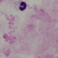
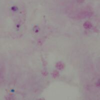
The parasites on a thick film lie within a background of white cells, platelets and various red cell components that have not been fully lysed. It is important to recognise these different features (which may vary a little between films depending on thickness and staining). A film area without parasites is shown below.
-
A
-
B


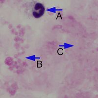
Normal background elements of a thick malaria film. The neutrophil (A) is recognisable by the characteristic nuclear shape but show artefactual distortion: in this case the chromatin detail is lost and there is no visible membrane outline or granular content. A careful look at the group of structures (B) suggests that they are part of a large group of clumped platelets formed when the film was prepared. Finally, the more amorphous material (C) most likely represents fibrin clot and un-lysed red cell debris.
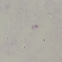
Precise appearances can vary and additional examples are shown below.
-
C
-

D


In the example C the red cells are incompletely lysed in a less thick area of the film and can be distinguished as separate cells, although with reduced haemoglobin content indicated by their yellow shade. The example D is similar to the previous images (A & B) but with better preservation and granulation of the neutrophil.
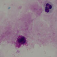
Distinguishing parasites on thick films
-
A
-

B


Subtle single parasite
-
C
-

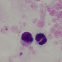
D


Two parasites with debris
-
A
-

B


3 parasites