Thick films - parasites identification
From MalariaETC
Navigation
>Main Malaria Index
>>Thick film - main page
>>>Current page: Distinguishing features on thick film
-

Low magnification view for scale, 3 regions marked
-

Region A: a single disrupted trophozoite
-
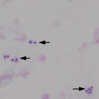
Region B: 5 trophozoites in three group
-
Region C: 2 trophozoites in one group


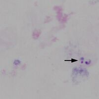
"
Note the differences in recognition - the typical ring form and vacuole of the parasite are not as easy to distinguish and chromatin dots may appear to separate from parasite cytoplasm while the absence of intact red cells takes away important clues to parasite size, distribution within the red cell, and any red cell changes. This is illustrated in the image below