Thick films - parasites identification: Difference between revisions
From MalariaETC
No edit summary |
No edit summary |
||
| Line 26: | Line 26: | ||
File:4 LT_PO_dots.jpg|B|link={{filepath:4 LT_PO_dots_anno.jpg}} | File:4 LT_PO_dots.jpg|B|link={{filepath:4 LT_PO_dots_anno.jpg}} | ||
</gallery> | </gallery> | ||
<span style="font-size:90%">These examples show slightly better preservation than the early trophozoites in the previos example. Further supporting the detection of "species-specific" parasite features. For example A (''P.vivax'') the parasite | <span style="font-size:90%">These examples show slightly better preservation than the early trophozoites in the previos example. Further supporting the detection of "species-specific" parasite features. For example A (''P.vivax'') the two visible parasite are large and almost fill the erythrocyte with irregularity of parasite form that distorts the ring shape; this accompanied by distortion of the shape of the containing erythrocyte and by the suggesion of Schuffner's dots in the red cell cytoplasm. The example cells in image B (''P.ovale'') also have large paraites with cytoplasmic dots in the erythrocyte, but with a relatively preserved ring form and round but fimbriated red cells.</span> | ||
---- | ---- | ||
<span style="font-size:90%">'''Schizont forms'''</span> | <span style="font-size:90%">'''Schizont forms'''</span> | ||
Revision as of 10:27, 4 March 2025
Navigation
>Main Malaria Index
>>Thick film - main page
>>>Current page: Parasite identification using thick films
Recognising specific parasite features on malaria thick films
In some species and parasite forms the morphology will be better preserved allowing a more confident identification and assignment of either parasite developmental stage, species or associated species. This can vary within the preparation according to the thickness of the preparation and the relative rsistance to lysis. Some examples are given below:
NOTE: the images are at relatively low magnification - to view them properly please click on the image to view in larger annotated form, then use the "back" button on your browser to return to the referring page.
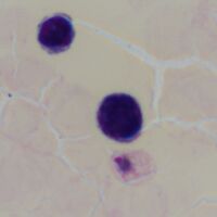
Early trophozoite forms (P.ovale)
-
A
-
B


The early trophozoites shown on this thick-film image are well preserved with a well-defined chromatin dot and cytoplasm; in contrast to many examples found on thick films the infected red cell has resisted complete lysis allowing its outline to be distinguished and also the frequent (although less well defined) James' dots to be seen (as well as a suggestion of gold/brown pigment). This makes it possible to infer the species as likely to be P.ovale or P.vivax although a thin film would provide much greater diagnostic confidence and is recommended for species identification.
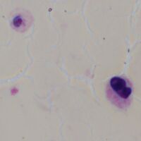
Late trophozoite forms (P.vivax and P.ovale)
-
A
-
B


These examples show slightly better preservation than the early trophozoites in the previos example. Further supporting the detection of "species-specific" parasite features. For example A (P.vivax) the two visible parasite are large and almost fill the erythrocyte with irregularity of parasite form that distorts the ring shape; this accompanied by distortion of the shape of the containing erythrocyte and by the suggesion of Schuffner's dots in the red cell cytoplasm. The example cells in image B (P.ovale) also have large paraites with cytoplasmic dots in the erythrocyte, but with a relatively preserved ring form and round but fimbriated red cells.
Schizont forms


Gametocyte forms


Malaria pigment

