Thick films - parasites and debris: Difference between revisions
From MalariaETC
No edit summary |
No edit summary |
||
| Line 31: | Line 31: | ||
File:12 Mess & troph anno.jpg|<span style="font-size:80%">'''B'''</span>|link={{filepath:12 Mess & troph anno.jpg}} | File:12 Mess & troph anno.jpg|<span style="font-size:80%">'''B'''</span>|link={{filepath:12 Mess & troph anno.jpg}} | ||
</gallery> | </gallery> | ||
<span style="font-size:90%">Subtle single parasite</span> | <span style="font-size:90%">Subtle single parasite</span> | ||
</br></br> | </br></br> | ||
| Line 38: | Line 37: | ||
File:13 Mess & troph anno.jpg|<span style="font-size:80%">'''D'''</span>|link={{filepath:13 Mess & troph anno.jpg}} | File:13 Mess & troph anno.jpg|<span style="font-size:80%">'''D'''</span>|link={{filepath:13 Mess & troph anno.jpg}} | ||
</gallery> | </gallery> | ||
<span style="font-size:90%">Two parasites with debris</span> | <span style="font-size:90%">Two parasites with debris</span> | ||
</br></br> | </br></br> | ||
| Line 45: | Line 43: | ||
File:14 Mess & troph anno.jpg|<span style="font-size:80%">'''B'''</span>|link={{filepath:14 Mess & troph anno.jpg}} | File:14 Mess & troph anno.jpg|<span style="font-size:80%">'''B'''</span>|link={{filepath:14 Mess & troph anno.jpg}} | ||
</gallery> | </gallery> | ||
<span style="font-size:90%">3 parasites</span> | <span style="font-size:90%">3 parasites</span> | ||
---- | ---- | ||
Revision as of 21:09, 21 February 2025
Navigation
>Main Malaria Index
>>Thick film - main page
>>>Current page: Distinguishing features on thick film
Recognising parasites on thick malaria films
Recognising "debris" on thick films
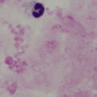
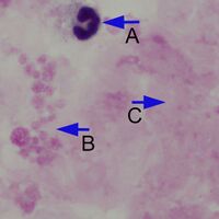
The parasites on a thick film lie within a background of white cells, platelets and various red cell components that have not been fully lysed. It is important to recognise these different features (which may vary a little between films depending on thickness and staining). A film area without parasites is shown below.
-
A
-
B


Normal background elements of a thick malaria film. The neutrophil (A) is recognisable by the characteristic nuclear shape but show artefactual distortion: in this case the chromatin detail is lost and there is no visible membrane outline or granular content. A careful look at the group of structures (B) suggests that they are part of a large group of clumped platelets formed when the film was prepared. Finally, the more amorphous material (C) most likely represents fibrin clot and un-lysed red cell debris.
Precise appearances can vary and additional examples are shown below.
-
C
-

D


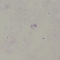
In the example C the red cells are incompletely lysed in a less thick area of the film and can be distinguished as separate cells, although with reduced haemoglobin content indicated by their yellow shade. The example D is similar to the previous images (A & B) but with better preservation and granulation of the neutrophil.
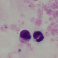
Distinguishing parasites on thick films
-
A
-

B


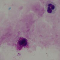
Subtle single parasite
-
C
-

D


Two parasites with debris
-
A
-

B


3 parasites