Thick films - parasites and debris: Difference between revisions
From MalariaETC
No edit summary |
No edit summary |
||
| Line 17: | Line 17: | ||
</br> | </br> | ||
<gallery mode="nolines" heights=200px widths=200px> | <gallery mode="nolines" heights=200px widths=200px> | ||
File:12 Mess & troph.jpg|<span style="font-size:80%"> | File:12 Mess & troph.jpg|<span style="font-size:80%">'''A'''</span>|link={{filepath:11 Mess only.jpg}} | ||
File:12 Mess & troph anno.jpg|<span style="font-size:80%"> | File:12 Mess & troph anno.jpg|<span style="font-size:80%">'''B'''</span>|link={{filepath:12 Mess & troph anno}} | ||
</gallery> | </gallery> | ||
</br> | </br> | ||
| Line 25: | Line 25: | ||
</br> | </br> | ||
<gallery mode="nolines" heights=200px widths=200px> | <gallery mode="nolines" heights=200px widths=200px> | ||
File:13 Mess & troph.jpg|<span style="font-size:80%"> | File:13 Mess & troph.jpg|<span style="font-size:80%">'''A'''</span>|link={{filepath:13 Mess & troph.jpg}} | ||
File:13 Mess & troph anno.jpg|<span style="font-size:80%"> | File:13 Mess & troph anno.jpg|<span style="font-size:80%">'''B'''</span>|link={{filepath:13 Mess & troph anno.jpg}} | ||
</gallery> | </gallery> | ||
</br> | </br> | ||
| Line 34: | Line 34: | ||
</br> | </br> | ||
<gallery mode="nolines" heights=200px widths=200px> | <gallery mode="nolines" heights=200px widths=200px> | ||
File:14 Mess & troph.jpg|<span style="font-size:80%"> | File:14 Mess & troph.jpg|<span style="font-size:80%">'''A'''</span>|link={{filepath:14 Mess & troph.jpg}} | ||
File:14 Mess & troph anno.jpg|<span style="font-size:80%"> | File:14 Mess & troph anno.jpg|<span style="font-size:80%">L'''B'''</span>|link={{filepath:14 Mess & troph anno.jpg}} | ||
</gallery> | </gallery> | ||
</br> | </br> | ||
<span style="font-size:90%">Note the differences in recognition - the typical ring form and vacuole of the parasite are not as easy to distinguish and chromatin dots may appear to separate from parasite cytoplasm while the absence of intact red cells takes away important clues to parasite size, distribution within the red cell, and any red cell changes. This is illustrated in the image below</span> | <span style="font-size:90%">Note the differences in recognition - the typical ring form and vacuole of the parasite are not as easy to distinguish and chromatin dots may appear to separate from parasite cytoplasm while the absence of intact red cells takes away important clues to parasite size, distribution within the red cell, and any red cell changes. This is illustrated in the image below</span> | ||
---- | ---- | ||
Revision as of 20:04, 14 February 2025
Navigation
>Main Malaria Index
>>Thick film - main page
>>>Current page: Distinguishing features on thick film
-
A
-
B


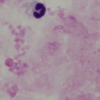
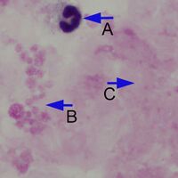
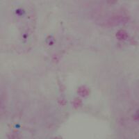
Note the differences in recognition - the typical ring form and vacuole of the parasite are not as easy to distinguish and chromatin dots may appear to separate from parasite cytoplasm while the absence of intact red cells takes away important clues to parasite size, distribution within the red cell, and any red cell changes. This is illustrated in the image below
-
A
-

B
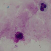


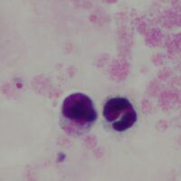
Note the differences in recognition - the typical ring form and vacuole of the parasite are not as easy to distinguish and chromatin dots may appear to separate from parasite cytoplasm while the absence of intact red cells takes away important clues to parasite size, distribution within the red cell, and any red cell changes. This is illustrated in the image below
-
A
-

LB


Note the differences in recognition - the typical ring form and vacuole of the parasite are not as easy to distinguish and chromatin dots may appear to separate from parasite cytoplasm while the absence of intact red cells takes away important clues to parasite size, distribution within the red cell, and any red cell changes. This is illustrated in the image below