Thick film interpretation: Difference between revisions
From MalariaETC
No edit summary |
No edit summary |
||
| Line 27: | Line 27: | ||
</br> | </br> | ||
'''Detailed Sections:'''</br></br> | '''Detailed Sections:'''</br></br> | ||
<span style="font-size:200%">→</span> [[Thick_vs_Thin_film_comparison| | |||
Section 1: a comparison of the strengths and weaknesses of the different morphological approaches to malaria diagnosis | |||
<span style="font-size:200%">→</span> [[Thick_vs_Thin_film_comparison|Click to see comparison of thick and thin film approaches]] | |||
COMPARISON THICK vs THIN</br> | COMPARISON THICK vs THIN</br> | ||
Revision as of 11:09, 13 February 2025
| OVERVIEW OF THICK FILMS |
A thick film is prepared by placing a small drop of blood on a slide then spreading it in a circular motion. The thick layer acheived is then air-dried without fixation.
IMAGE
The principles are:
- The blood layer will be many layers thick (varying from 6-20 accross the specimen)
- The erythrocytes are unfixed, so will be lysed during staining appearing only as debris.
- The Giemsa stain will stain and distinguish the remaining white cells and parasites.
- This concentration effect allows parasites to be detected with high sensitivity
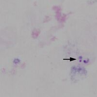
Typical appearances of a case of P.falciparum with easily detected trophozoites are shown below.
-

Low magnification view for scale, 3 regions marked
-

Region A: a single disrupted trophozoite
-
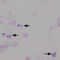
Region B: 5 trophozoites in three group
-
Region C: 2 trophozoites in one group


"
Note the differences in recognition - the typical ring form and vacuole of the parasite are not as easy to distinguish and chromatin dots may appear to separate from parasite cytoplasm while the absence of intact red cells takes away important clues to parasite size, distribution within the red cell, and any red cell changes. This is illustrated in the image below
-

Two ring frms of P.falciparum
-
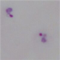
High-power image of the parasites


Detailed Sections:
Section 1: a comparison of the strengths and weaknesses of the different morphological approaches to malaria diagnosis → Click to see comparison of thick and thin film approaches
COMPARISON THICK vs THIN
DISTINGUISHING PARASITES vs DEBRIS
DISTINGUISHING SPECIES
IDENTIFYING PIGMENT
Some strengths and weaknesses of each approach are summarised in the table below: