Thick film interpretation: Difference between revisions
From MalariaETC
No edit summary |
No edit summary |
||
| Line 3: | Line 3: | ||
|colspan="1" style = "font-size:100%; color:black; background: gainsboro |'''OVERVIEW OF THICK FILMS''' | |colspan="1" style = "font-size:100%; color:black; background: gainsboro |'''OVERVIEW OF THICK FILMS''' | ||
|} | |} | ||
</br> | |||
IMAGE | |||
</br> | </br> | ||
<span style="font-size:90%">A thick film is prepared by placing a small drop of blood on a slide then spreading it in a circular motion. The thick layer acheived is then air-dried without fixation.</br></br>The principles are:</br> | <span style="font-size:90%">A thick film is prepared by placing a small drop of blood on a slide then spreading it in a circular motion. The thick layer acheived is then air-dried without fixation.</br></br>The principles are:</br> | ||
Revision as of 19:52, 10 February 2025
| OVERVIEW OF THICK FILMS |
IMAGE
A thick film is prepared by placing a small drop of blood on a slide then spreading it in a circular motion. The thick layer acheived is then air-dried without fixation.
The principles are:
- The blood will therefore be many layers thick (around 6-20) compared with the single layer of a thin film
- The erythrocytes are unfixed so will be lysed during staining appearning only as debris.
- The Giemsa stain will therefore stain and distinguish the remaining white cells, parasites.
- This allows parasites to be detected with high sensitivity using fewer microscopic fields
The features described make this approach highly sensitive for parasite detection, but also introduces staining inconsistencies and possible disruption of parasites, analysis therefore requires experienced microscopists who are aware of protential artefacts and have experience in thick blood film interpretation. Typial appearnces of a case of P.falciparum with easily detected trophozoites is shown below.
-

Low magnification view for scale, 3 regions marked
-

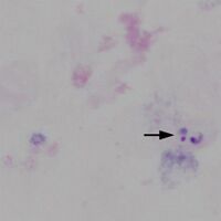
Region A: a single disrupted trophozoite
-
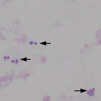
Region B: 5 trophozoites in three group
-
Region C: 2 trophozoites in one group


"
| COMPARISON OF THICK AND THIN FILMS |
| Feature | Thick Film | Thin Film |
|---|---|---|
| Sensitivity for detection | Higher: detects low parasitaemia ~5–10 parasites/µL | Lower: generally needs ~50 parasites/µL for reliable detection) |
| Species Identification | Poor: RBC morphology lost and species-specific features may be difficult | Excellent: Parasite morphology and RBC characteristics are readily observed |
| Quantification of parasitaemia | Difficult: requires estimation so is imprecise | Easier: parasites can be counted per number of RBCs |
| Preparation and staining | Longer: requires air drying before careful staining to avoid artefact | Faster: films are fixed and stained immediately with clearer morphology |