Gallery of early trophozoites: Difference between revisions
From MalariaETC
No edit summary |
No edit summary |
||
| (23 intermediate revisions by the same user not shown) | |||
| Line 3: | Line 3: | ||
'''Navigation'''</br> | '''Navigation'''</br> | ||
<span style="font-size:90%">>[[MalariaETC_Index|Main Malaria Index]]''</span></br> | <span style="font-size:90%">>[[MalariaETC_Index|Main Malaria Index]]''</span></br> | ||
<span style="font-size:90%">>>[[ | <span style="font-size:90%">>>[[Galleries|Galleries Index Page]]''</span></br> | ||
<span style="font-size:90%">>>>Current page: ''' | <span style="font-size:90%">>>>Current page: '''Gallery of early trophozoites'''</span> | ||
---- | ---- | ||
<span style="font-size: | <span style="font-size:140%; color:navy">'''Gallery of Early Trophozoites'''</br></span> | ||
</br> | |||
<span style="font-size: | <span style="font-size:90%">At this very earliest developmental stage all trophozoites appear as ring forms. In the earliest forms any differences between species rely most on size and thickness such that species differences are very difficult to distinguish - however in many cases recognisable "species specific" features begin to appear as parasites mature toward late trophozoite stages.</br></br> | ||
---- | ---- | ||
<span style="font-size:90%">''' ''P.falciparum'' '''</span></br> | <span style="font-size:90%">''' ''P.falciparum'' '''</span></br> | ||
<span style="font-size:90%">Small delicate rings, and | <span style="font-size:90%">Small delicate rings, and may be the only forms seen in some patients at diagnosis. The infected red cells have normal (or slightly crenated) appearance.</br> | ||
<gallery mode="nolines" heights=200px widths=200px> | |||
<gallery heights=200px widths=200px> | |||
File:PFET1g.jpg|<span style="font-size:80%">Fine ring form</span>|link={{filepath:PFET1g.jpg}} | File:PFET1g.jpg|<span style="font-size:80%">Fine ring form</span>|link={{filepath:PFET1g.jpg}} | ||
File:PFET2g.jpg|<span style="font-size:80%">Double dot form and normal ring</span>|link={{filepath:PFET2g.jpg}} | File:PFET2g.jpg|<span style="font-size:80%">Double dot form and normal ring</span>|link={{filepath:PFET2g.jpg}} | ||
File:PFET3g.jpg|<span style="font-size:80%">Accolé and double dot forms</span>|link={{filepath:PFET3g.jpg}} | File:PFET3g.jpg|<span style="font-size:80%">Accolé and double dot forms</span>|link={{filepath:PFET3g.jpg}} | ||
File:PFET4g.jpg|<span style="font-size:80%">Multiple parasite form</span>|link={{filepath:PFET4g.jpg}} | File:PFET4g.jpg|<span style="font-size:80%">Multiple parasite form</span>|link={{filepath:PFET4g.jpg}}</gallery>" | ||
</gallery>" | |||
---- | ---- | ||
<span style="font-size:95%">''' ''P.vivax'' '''</span></br> | <span style="font-size:95%">''' ''P.vivax'' '''</span></br> | ||
<span style="font-size:90%">Rings begin as small forms, but become larger asociated with enlarged distorted red cells as they develop. Schüffner's | <span style="font-size:90%">Rings begin as small forms, but become larger asociated with enlarged distorted red cells as they develop. Schüffner's dots will appear during this stage. | ||
<gallery heights=200px widths=200px> | <gallery mode="nolines" heights=200px widths=200px> | ||
File: | File:PVET1.jpg|<span style="font-size:80%">Early ring form</span>|link={{filepath:PVET1.jpg}} | ||
File: | File:PVET2.jpg|<span style="font-size:80%">Early ring form with faint dots</span>|link={{filepath:PVET2.jpg}} | ||
File: | File:PVET3.jpg|<span style="font-size:80%">Large thickened ring trophozoite</span>|link={{filepath:PVET3.jpg}} | ||
File: | File:PVET4.jpg|<span style="font-size:80%">Ring/amoeboid trophozoites, Schüffner's dots</span>|link={{filepath:PVET4.jpg}} | ||
</gallery>" | </gallery>" | ||
---- | ---- | ||
<span style="font-size:90%">''' ''P.ovale'' '''</span></br> | <span style="font-size:90%">''' ''P.ovale'' '''</span></br> | ||
<span style="font-size:90%"> | <span style="font-size:90%">The "ring" shape is retained but becomes larger and thicker, red cells may develop fimbriation and enlarged ovoid form with visible James' dots as development progresses. | ||
<gallery mode="nolines" heights=200px widths=200px> | |||
<gallery mode=" | File:POET1.jpg|<span style="font-size:80%">Early ring form</span>|link={{filepath:POET1.jpg}} | ||
File: | File:POET2.jpg|<span style="font-size:80%">Ring with dots/fimbriation</span>|link={{filepath:POET2.jpg}} | ||
File: | File:POET3.jpg|<span style="font-size:80%">faint James' dots</span>|link={{filepath:POET3.jpg}} | ||
File: | File:POET4.jpg|<span style="font-size:80%">Ring early ovoid change</span>|link={{filepath:POET4.jpg}}</gallery>" | ||
File: | |||
</gallery>" | |||
---- | ---- | ||
<span style="font-size:90%">''' ''P.malariae'' '''</span></br> | <span style="font-size:90%">''' ''P.malariae'' '''</span></br> | ||
<span style="font-size:90%">Infected red cells are generally infrequent. Early trophozoites are small in normal or small erythrocytes, and may have central chromatin dot, elongation or angular forms. | <span style="font-size:90%">Infected red cells are generally infrequent. Early trophozoites are small in normal or small erythrocytes, and may have central chromatin dot, elongation or angular forms. | ||
<gallery mode="nolines" heights=200px widths=200px> | |||
<gallery mode=" | |||
File:MET1g.jpg|<span style="font-size:80%">Ring form in small red cell</span>|link={{filepath:MET1g.jpg}} | File:MET1g.jpg|<span style="font-size:80%">Ring form in small red cell</span>|link={{filepath:MET1g.jpg}} | ||
File:MET2g.jpg|<span style="font-size:80%">The central chromatin dot</span>|link={{filepath:MET2g.jpg}} | File:MET2g.jpg|<span style="font-size:80%">The central chromatin dot</span>|link={{filepath:MET2g.jpg}} | ||
File:PMET3g.jpg|<span style="font-size:80%">Early elongation, | File:PMET3g.jpg|<span style="font-size:80%">Early elongation, Ziemann's dots</span>|link={{filepath:PMET3g.jpg}} | ||
File:MET4g.jpg|<span style="font-size:80%">Early angularity of form</span>|link={{filepath:MET4g.jpg}} | File:MET4g.jpg|<span style="font-size:80%">Early angularity of form</span>|link={{filepath:MET4g.jpg}} | ||
</gallery>" | </gallery>" | ||
---- | ---- | ||
<span style="font-size:90%">''' ''P.knowlesi'' '''</span></br> | <span style="font-size:90%">''' ''P.knowlesi'' '''</span></br> | ||
<span style="font-size:90%">The early trophozoite may | <span style="font-size:90%">The early trophozoite may resemble ''P.falciparum'' and infected cells may be frequent. Later forms however begin to resemble parasites of ''P.malariae''. | ||
<gallery mode="nolines" heights=200px widths=200px> | |||
<gallery mode=" | File:PKET1a.jpg|<span style="font-size:80%">Fine early rings</span>|link={{filepath:PKET1a.jpg}} | ||
File: | |||
File:PKET2a.jpg|<span style="font-size:80%">Double dot (right)</span>|link={{filepath:PKET2a.jpg}} | File:PKET2a.jpg|<span style="font-size:80%">Double dot (right)</span>|link={{filepath:PKET2a.jpg}} | ||
File:PKET3a.jpg|<span style="font-size:80%">Accolé form</span>|link={{filepath:PKET3a.jpg}} | File:PKET3a.jpg|<span style="font-size:80%">Accolé form</span>|link={{filepath:PKET3a.jpg}} | ||
Latest revision as of 13:23, 17 March 2025
Navigation
>Main Malaria Index
>>Galleries Index Page
>>>Current page: Gallery of early trophozoites
Gallery of Early Trophozoites
At this very earliest developmental stage all trophozoites appear as ring forms. In the earliest forms any differences between species rely most on size and thickness such that species differences are very difficult to distinguish - however in many cases recognisable "species specific" features begin to appear as parasites mature toward late trophozoite stages.
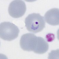
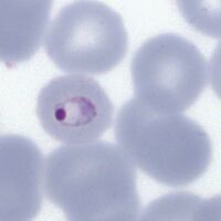
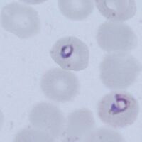
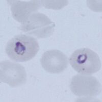
P.falciparum
Small delicate rings, and may be the only forms seen in some patients at diagnosis. The infected red cells have normal (or slightly crenated) appearance.
-
Fine ring form
-
Double dot form and normal ring
-
Accolé and double dot forms
-
Multiple parasite form


"
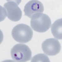
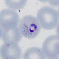
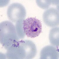
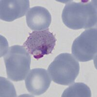
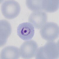
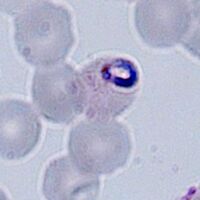
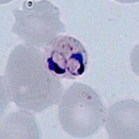
P.vivax
Rings begin as small forms, but become larger asociated with enlarged distorted red cells as they develop. Schüffner's dots will appear during this stage.
-
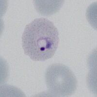
Early ring form
-
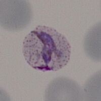
Early ring form with faint dots
-
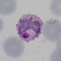
Large thickened ring trophozoite
-
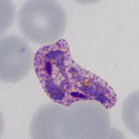
Ring/amoeboid trophozoites, Schüffner's dots


"
P.ovale
The "ring" shape is retained but becomes larger and thicker, red cells may develop fimbriation and enlarged ovoid form with visible James' dots as development progresses.
-
Early ring form
-
Ring with dots/fimbriation
-
faint James' dots
-
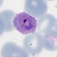
Ring early ovoid change


"
P.malariae
Infected red cells are generally infrequent. Early trophozoites are small in normal or small erythrocytes, and may have central chromatin dot, elongation or angular forms.
-
Ring form in small red cell
-
The central chromatin dot
-
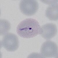
Early elongation, Ziemann's dots
-
Early angularity of form


"
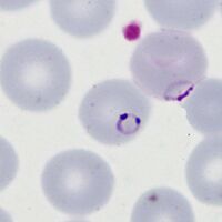
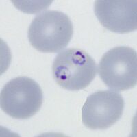
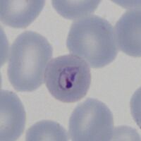
P.knowlesi
The early trophozoite may resemble P.falciparum and infected cells may be frequent. Later forms however begin to resemble parasites of P.malariae.
-
Fine early rings
-
Double dot (right)
-
Accolé form
-
Multiple infection


