Gallery of schizonts
From MalariaETC
Navigation
>Main Malaria Index
>>Galleries Index Page
>>>Current page: Gallery of schizonts
Gallery of Schizonts
Schizont morphology is variable as they progress from late trophoxoites, dividing their chromatin into seprate distict masses (usually a schizont is defined by have more than two masses to distinguish them from rings with double dots). The morphological variability then continues as the merozoites separate before release. However, despite this, some features such as erythocyte size and shape, added dots, pigment distribution and the number of merozoites present can still be useful (as can the fact that they are rarely seen in P.falciparum.
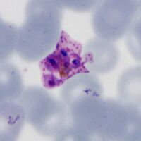
P.falciparum
Loose and often "tatty" appearances with 8-16 merozoites and clumped pigment when mature. Rare in blood as they sequesterin tissues and circulating form may appear degenerate.
-
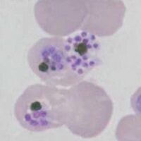
Variable merozoite number, clumped pigment
-
Degenerate small merozoite
-

Large merozoites
-
Large merozoites


"
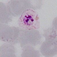
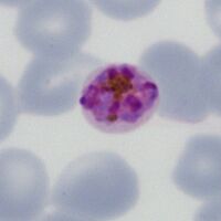
P.vivax
Charactertically large and red cell dots may be seen. The merozoites tend to pack the red cell with numbers up to 16-32 in mature merozoites.
-

Large very irregular ring, Schüffner's dots
-

Amoeboid parasite, distorted red cell
-

Aomeboid parasite, clumped pigment
-

Very large amoeboid form


"
P.ovale
Share many features with P.vivax and may not be easy to distinguish, tend not to be as large with up to 16 merozoites. Ovoid shape and fimbriation of red cells may be present.
-
Ring form retained, fimbriationa and dots
-
Ring form retained, ovoid red cell
-

Solid parasite, red cell fimbriation ("comet form")
-

Solid parasite, red cell fimbriation


"
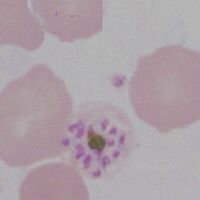
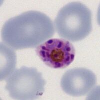
P.malariae
Infected red cells may be infrequent. Parasites may become more solid and angular, or become elonagated and may extend across the red cell (band appearance). Red cells remain round and may be small, added dots (Ziemann's dots) are rarely seen.
-

Early elongated form, small red cell
-
A full band across the red cells
-
An open angular parasite, note pigment
-
Angularity form


"
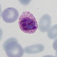
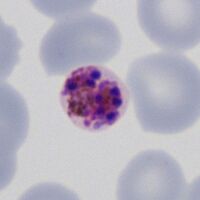
P.knowlesi
Late trophozoite forms may still resemble P.falciparum but also develop features of P.malariae, although number may be high and dots (Sinton and Mulligan's stippling) are more likely to be seen.
-

Mixed irregular rings sparse dots
-

Elongated parasite forms
-

Solid and angular forms
-

Mixed forms, dots and pigment


