Gallery of late trophozoites
From MalariaETC
Navigation
>Main Malaria Index
>>Galleries Index Page
>>>Current page: Gallery of late trophozoites
Gallery of Late Trophozoites
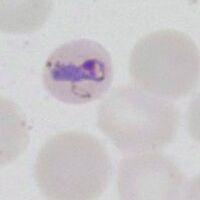
Late trophozoites often have "species-specific" changes that affect parasite, red cell shape or added dots. These may be very helpful in assigning diagnosis, although not all changes are fully specific to a single species.
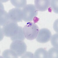
P.falciparum
The key features here are a slight thickening of ring forms but with the same appearances as early trophozoites. Additionally look for Maurer's dots and clefts appearning.
-

Two parasites, Maurer's dots
-

Double dot, Maurer's dots
-
Accolé and Maurer's dots
-

Accolé and Maurer's dots


"
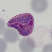
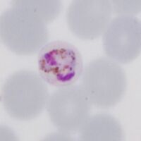
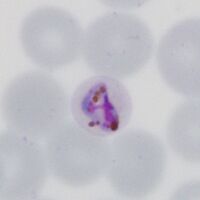
P.vivax
Rings begin as small forms, but become larger asociated with enlarged distorted red cells as they develop. Schüffner's dots will become present
-
Early ring form
-
Early ring form with faint dots
-

Llarge thickened ring trophozoite
-

Ring trophozoites, Schüffner's dots


"
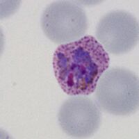
P.ovale
Ring form is retained but enlarges, red cells may develop fimbriation and enlarged ovoid form with visible James' dots.
-

Early ring form
-

Ring with dots/fimbriation
-
faint Ziemann's dots
-

Ring early ovoid change


"
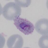
P.malariae
Infected red cells are generally infrequent. Early trophozoites are small in normal or small erythrocytes, and may have central chromatin dot, elongation or angular forms.
-
Ring form in small red cell
-

The central chromatin dot
-
Early elongation, Stinton's dots
-
Early angularity of form


"
P.knowlesi
The early trophozoite may resembles P.falciparum and infected cells may be frequent. Later forms however begin to resemble parasites of P.malariae.
-

Fine early rings
-

Double dot (right)
-

Accolé form
-

Multiple infection


