Gallery of late trophozoites
From MalariaETC
Navigation
>Main Malaria Index
>>Galleries Index Page
>>>Current page: Gallery of late trophozoites
Gallery of Late Trophozoites
Late trophozoites often have "species-specific" changes that affect parasite, red cell shape or added dots. These may be very helpful in assigning diagnosis, although not all changes are fully specific to a single species.
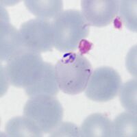
P.falciparum
The key features here are a slight thickening of ring forms but with the same appearances as early trophozoites. Additionally look for Maurer's dots and clefts appearning.
-

Two parasites, Maurer's dots and clefts
-

Double dot form with Maurer's dots
-
Accolé and double dot forms
-

Multiple parasite form


"
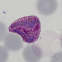
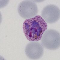
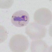
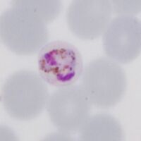
P.vivax
Ring forms are replaced with irregular and "amoeboid" forms. Red cells and parasites become markedly larger with distortion of red cells as they develop. Schüffner's dots and pigment becomes prominent.
-
Early ring form
-
Early ring form with faint dots
-

Llarge thickened ring trophozoite
-

Ring trophozoites, Schüffner's dots


"
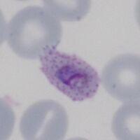
P.ovale
Ring form is retained but enlarges, red cells may develop fimbriation and enlarged ovoid form with visible James' dots.
-

Early ring form
-

Ring with dots/fimbriation
-
faint Ziemann's dots
-

Ring early ovoid change


"
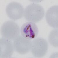
P.malariae
Infected red cells are generally infrequent. Early trophozoites are small in normal or small erythrocytes, and may have central chromatin dot, elongation or angular forms.
-
Ring form in small red cell
-

The central chromatin dot
-
Early elongation, Stinton's dots
-
Early angularity of form


"
P.knowlesi
The early trophozoite may resembles P.falciparum and infected cells may be frequent. Later forms however begin to resemble parasites of P.malariae.
-

Fine early rings
-

Double dot (right)
-

Accolé form
-

Multiple infection


