Plasmodium vivax: Morphology: Difference between revisions
From MalariaETC
No edit summary |
No edit summary |
||
| Line 37: | Line 37: | ||
</br><span style="font-size:130%">The late trophozoite</span> | </br><span style="font-size:130%">The late trophozoite</span> | ||
<gallery mode="nolines" widths=250px heights=250px> | <gallery mode="nolines" widths=250px heights=250px> | ||
File: | File:PVLTc.jpg|link={{filepath:PVLTc.jpg}} | ||
File: | File:PVLT1.jpg|link={{filepath:PVLT1p.jpg}} | ||
</gallery> | </gallery> | ||
<br clear=all> | <br clear=all> | ||
The later growth stage | The later growth stage during which parasites grow considerably and lose their ring appearance, this process is accompanied by substantial modification of the red cell and metabolism of it's haemoglobin to form malaria pigment. | ||
* | *infected erythrocytes become significantly enlarged and irregular in shape | ||
* | *parasites lose their ring appearnace becoming irregular and "[[amoeboid]]" in form | ||
* | *numerous red/purple Schüffner's dots are predent in the cytoplasm of red cells | ||
*[[malaria pigment]] is often present and has an irregular distribution | |||
* | |||
</br> | </br> | ||
<div style="width: 350px"> | <div style="width: 350px"> | ||
Revision as of 17:39, 11 December 2024
| Navigation |
| >Main malaria Index |
| >Main Species identification page |
Morphology of Plasmodium vivax
(See Malaria Biology pages for an explanation of these stages)
The early trophozoite


The earliest ring forms may be indistinguishable from other species, but during this stage the parasite tends to aquire a more irregular forms and to show signs of modification of the erythrocyte (added dots, and altered size and shape).
- erythrocytes begin to show increased size and altered shape
- parasites retain a ring form but may aquire a more irregular form
- parasites are generally large - occupying up to half of the erythrocyte
- cytoplasmic Schüffner's dots may appear at this stage, although pigment is less uncommon
The late trophozoite

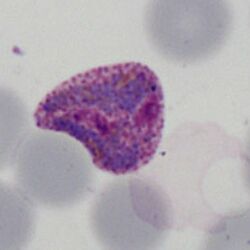
The later growth stage during which parasites grow considerably and lose their ring appearance, this process is accompanied by substantial modification of the red cell and metabolism of it's haemoglobin to form malaria pigment.
- infected erythrocytes become significantly enlarged and irregular in shape
- parasites lose their ring appearnace becoming irregular and "amoeboid" in form
- numerous red/purple Schüffner's dots are predent in the cytoplasm of red cells
- malaria pigment is often present and has an irregular distribution
The schizont


The schizont is the asexual form of the malaria parasite that will circulate in blood in most species, but is an uncommon feature in P.falciparum:
- Do not generally circulate in this species unless overwhelming infection
- The merozoites cluster "untidily" but may be numerous (8-16+ when mature)
- In this species the loose pigment may be seen in clumps between the parasites
- Red cell size is generally unaffected but red cells become pale as haemoglobin is metabolised by the parasites
The gametocyte


The sexual replication in P.falciparum is very distinctive and may be the only form visible (particularly of after treatment).
- male and femaie gametocytes have the appearance of rods although these may be distorted
- The rod shapes may become curved by the red cell membrane to give the characteristic "banana" form”
- The residual membrane (empty of haemoglobin) is often seen as a "blister" to one or both sides of the parasite
- The single chromatin area is in the centre of the parasite, often has pigment overlying it