Size and shape index: Difference between revisions
From MalariaETC
(Created page with "{{DISPLAYTITLE:<span style="position: absolute; clip: rect(1px 1px 1px 1px); clip: rect(1px, 1px, 1px, 1px);">{{FULLPAGENAME}}</span>}} '''Navigation'''</br> <span style="font-size:90%">>Main Malaria Index''</span></br> <span style="font-size:90%">>>Malaria Biology Index''</span></br> <span style="font-size:90%">>>>Biology of the trophozoite</span></br> <span style="font-size:90%">>>>Current p...") |
No edit summary |
||
| Line 1: | Line 1: | ||
{{DISPLAYTITLE:<span style="position: absolute; clip: rect(1px 1px 1px 1px); clip: rect(1px, 1px, 1px, 1px);">{{FULLPAGENAME}}</span>}} | {{DISPLAYTITLE:<span style="position: absolute; clip: rect(1px 1px 1px 1px); clip: rect(1px, 1px, 1px, 1px);">{{FULLPAGENAME}}</span>}} | ||
'''Navigation'''</br> | ---- | ||
{| class="wikitable" style="width: 60%; border-style: none; border-width: 0px; border-color: gainsboro; color:black" | |||
|style = "font-size:110%; color:black; background: gainsboro |'''Navigation'''</br> | |||
|- | |||
|<span style="font-size:110%">>[[Index|Previous Page]]''</span></br> | |||
|- | |||
|} | |||
---- | |||
{| class="wikitable" style="widthe:90%; border-style: solid; border-width: 4px; border-color:teal" | |||
|colspan="1" style = "font-size:140%; color:black; background: FFFAFA"|<span style="color:black>'''Cytoplasmic dots'''</span> | |||
---- | |||
<span style="font-size:90%"> | <span style="font-size:90%">The dots of different species represent parasite proteins that modify red cell function in a range of ways to support parasite development, immun evasion or virulence. Their precise functions are ill defined, but their appearances have value in distinguishing the different species of parasite. The dots require a well-stained specimen to be easily seen!</span> | ||
<span style="font-size:90% | ---- | ||
<span style="font-size:90% | <span style="font-size:90%>'''Schüffner's dots''' (P.vivax) and '''James' dots)''' (''P.ovale'') become apparent during the early trophzoite as faint dots then to be easily seen in later stages. These two forms of dots are morphologically indistinguishable as frequent evenly-distrubuted round dots od similar size.</span> | ||
< | <gallery mode="nolines" widths="220px" heights="220px" > | ||
File:Schuffner_dots.jpg|link={{filepath:Schuffner_dots.jpg}} | |||
File:James_dots.jpg|link={{filepath:James_dots.jpg}} | |||
</gallery> | |||
---- | |||
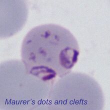
<span style="font-size:90%>'''Maurer's dots and clefts''' (''P.falciparum'') are blue/purple coloured and are not acquired until the late trophozoite stage of parasite development. They are less consistently "dot-like" and may appear as dots, clefts or plaques. '''Sinton and Mulligan's stippling''' (''P.knowlesi'') may appear similar.</span> | |||
<gallery mode="nolines" widths="220px" heights="220px" > | |||
File:Maurer_dots.jpg|link={{filepath:Maurer_dots.jpg.jpg}} | |||
File:Sinton_Mulligan_dots.jpg|link={{filepath:Sinton_Mulligan_dots.jpg}} | |||
</gallery> | |||
---- | ---- | ||
<span style="font-size:90%>'''Ziemann's stippling''' (''P.malariae'') Is not usully seen, but when present appears as faint fine (and inconspicuous) dots in cytoplasm of some ifected erythrocytes.</span> | |||
<gallery mode="nolines" widths="220px" heights="220px" > | |||
File:Ziemann_dots.jpg|link={{filepath:Ziemann_dots.jpg}} | |||
</gallery> | |||
{| class="wikitable" style="widthe:90%; border-style: solid; border-width: 4px; border-color:teal" | {| class="wikitable" style="widthe:90%; border-style: solid; border-width: 4px; border-color:teal" | ||
Revision as of 11:46, 10 December 2024
| Navigation |
| >Previous Page |
| Cytoplasmic dots
The dots of different species represent parasite proteins that modify red cell function in a range of ways to support parasite development, immun evasion or virulence. Their precise functions are ill defined, but their appearances have value in distinguishing the different species of parasite. The dots require a well-stained specimen to be easily seen! Schüffner's dots (P.vivax) and James' dots) (P.ovale) become apparent during the early trophzoite as faint dots then to be easily seen in later stages. These two forms of dots are morphologically indistinguishable as frequent evenly-distrubuted round dots od similar size. Maurer's dots and clefts (P.falciparum) are blue/purple coloured and are not acquired until the late trophozoite stage of parasite development. They are less consistently "dot-like" and may appear as dots, clefts or plaques. Sinton and Mulligan's stippling (P.knowlesi) may appear similar. Ziemann's stippling (P.malariae) Is not usully seen, but when present appears as faint fine (and inconspicuous) dots in cytoplasm of some ifected erythrocytes.
|