Gallery of schizonts: Difference between revisions
From MalariaETC
No edit summary |
No edit summary |
||
| Line 9: | Line 9: | ||
<span style="font-size:140%; color:navy">'''Gallery of Schizonts'''</br></span> | <span style="font-size:140%; color:navy">'''Gallery of Schizonts'''</br></span> | ||
</br> | </br> | ||
<span style="font-size:90%">Schizont morphology is variable as they progress from late trophoxoites, dividing their chromatin into seprate distict masses (usually a schizont is defined by have more than two masses to distinguish them from rings with double dots). The morphological variability then continues as the merozoites separate before release.</br></br> | <span style="font-size:90%">Schizont morphology is variable as they progress from late trophoxoites, dividing their chromatin into seprate distict masses (usually a schizont is defined by have more than two masses to distinguish them from rings with double dots). The morphological variability then continues as the merozoites separate before release. However, despite this, some features such as erythocyte size and shape, added dots, pigment distribution and the number of merozoites present can still be useful (as can the fact that they are rarely seen in ''P.falciparum''.</br></br> | ||
---- | ---- | ||
<span style="font-size:90%">''' ''P.falciparum'' '''</span></br> | <span style="font-size:90%">''' ''P.falciparum'' '''</span></br> | ||
Revision as of 21:19, 28 November 2024
Navigation
>Main Malaria Index
>>Galleries Index Page
>>>Current page: Gallery of schizonts
Gallery of Schizonts
Schizont morphology is variable as they progress from late trophoxoites, dividing their chromatin into seprate distict masses (usually a schizont is defined by have more than two masses to distinguish them from rings with double dots). The morphological variability then continues as the merozoites separate before release. However, despite this, some features such as erythocyte size and shape, added dots, pigment distribution and the number of merozoites present can still be useful (as can the fact that they are rarely seen in P.falciparum.
P.falciparum
The key features here are a slight thickening of ring forms but with the same appearances as early trophozoites. Additionally look for Maurer's dots and clefts appearning.
-
Two parasites, Maurer's dots and clefts
-
Double dot form with Maurer's dots
-

Accolé and double dot forms
-
Multiple parasite form


"
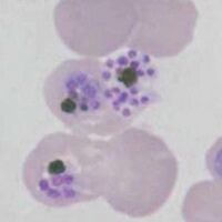
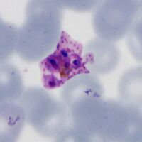
P.vivax
Ring forms are replaced with irregular and "amoeboid" forms. Red cells and parasites become markedly larger with distortion of red cells as they develop. Schüffner's dots and pigment becomes prominent.
-

Large very irregular ring, Schüffner's dots
-

Amoeboid parasite, distorted red cell
-

Aomeboid parasite, clumped pigment
-

Very large amoeboid form


"
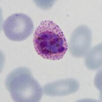
P.ovale
Cells and parasite enlarge, but ring form is often retained, red cells are a little enlarged with ovoid form and prominent James' dots.
-
Ring form retained, fimbriationa and dots
-
Ring form retained, ovoid red cell
-

Solid parasite, red cell fimbriation ("comet form")
-

Solid parasite, red cell fimbriation


"
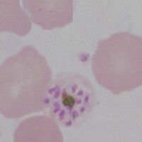
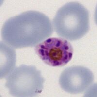
P.malariae
Infected red cells may be infrequent. Parasites may become more solid and angular, or become elonagated and may extend across the red cell (band appearance). Red cells remain round and may be small, added dots (Ziemann's dots) are rarely seen.
-

Early elongated form, small red cell
-
A full band across the red cells
-
An open angular parasite, note pigment
-
Angularity form


"
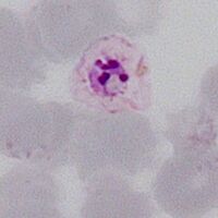
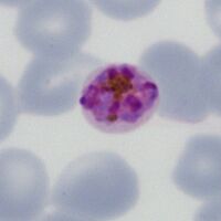
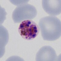
P.knowlesi
Late trophozoite forms may still resemble P.falciparum but also develop features of P.malariae, although number may be high and dots (Sinton and Mulligan's stippling) are more likely to be seen.
-

Mixed irregular rings sparse dots
-

Elongated parasite forms
-

Solid and angular forms
-

Mixed forms, dots and pigment


