RDT test: design and function: Difference between revisions
From MalariaETC
No edit summary |
No edit summary |
||
| Line 30: | Line 30: | ||
(1) In a typical test lysis buffer is loaded into one window ('''A'''), then the blood sample in a second window ('''B'''). Following this the lysed blood sample diffuses along the strip encontering lablelled antibody ('''Ab''') then continues to diffuse.</br></br> | (1) In a typical test lysis buffer is loaded into one window ('''A'''), then the blood sample in a second window ('''B'''). Following this the lysed blood sample diffuses along the strip encontering lablelled antibody ('''Ab''') then continues to diffuse into the window (arrow).</br></br> | ||
<gallery mode="nolines" widths=200px heights=300px> | <gallery mode="nolines" widths=200px heights=300px> | ||
Revision as of 08:31, 26 June 2024
Navigation
Go back
| Basic Principle |
If a patient has a malaria infection:
(1) A blood sample will be lysed in buffer to release parasite antigens, and expose them to dye-labelled antibodies
(2) The labelled antibodies will bind to parasite proteins forming an antigen-antibody complexes.
(3) These complexes are then captured by a second antibody immoblised on a nitro-cellulase to form a visible band.
(4) A control line binds lablelled antibody that has not bound malaria antigen showing the test was correctly performed

A positive "single band" type malaria test.
| How the tests work |
Test formats vary, but share common principles - this is illustrated for a single parasite antigen below.
(1) In a typical test lysis buffer is loaded into one window (A), then the blood sample in a second window (B). Following this the lysed blood sample diffuses along the strip encontering lablelled antibody (Ab) then continues to diffuse into the window (arrow).
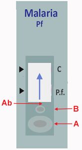
(2) The lysed blood and parasite antigens then meet a parasite-specific antibody labelled with dye (show strip and mixing of agents = A).

(2) Where the parasite-antigen is present, the dye-labelled antibody will bind to that antigen to form a complex (binding=B). This dye-labelled antigen/antibody complex then diffuses along the strip until it encounters a second parasite-specific antibody immobilised as a band on the strip. This immobilised antibody “captures” the labelled antibody/antigen complex to form a visible line (capture 1 = C).
(3) The remaining lysed sample (containing labelled antibody not bound to parasite antigen) continues to diffuse along the strip. And encounters a further immobilised antibody that captures it. This forms a control line which indicates that test has been successfully performed (capture 2 = D).