Multiple parasites: Difference between revisions
From MalariaETC
No edit summary Tag: Manual revert |
No edit summary |
||
| Line 4: | Line 4: | ||
|style = "font-size:110%; color:black; background: gainsboro |'''Navigation'''</br> | |style = "font-size:110%; color:black; background: gainsboro |'''Navigation'''</br> | ||
|- | |- | ||
|<span style="font-size:110%">>[[Plasmodium_falciparum:_Morphology| | |<span style="font-size:110%">>[[Plasmodium_falciparum:_Morphology|Return to previous Page]]''</span></br> | ||
|- | |- | ||
|} | |} | ||
| Line 36: | Line 36: | ||
<span style="font-size:90%">''Double parasites in: late trophozoite of P.malaria (A) late trophozoite of P.vivax (B) and late trophozoite of P.ovale (C)''</span> | <span style="font-size:90%">''Double parasites in: late trophozoite of P.malaria (A) late trophozoite of P.vivax (B) and late trophozoite of P.ovale (C)''</span> | ||
---- | |||
Revision as of 17:46, 26 December 2024
| Navigation |
| >Return to previous Page |
In some cases more than one parasite (most often though not always early or late trophozoites) infect a single erythrocyte. This is a surprisingly frequent finding, and it has been suggested some red cells are more attractive to parasites, or that already infected cells are more susceptible.

The most frequent form - two early trophozoites of P.falciparum in a single erythrocyte
Species significance
This is most often considered a feature indicating P.falciparum infection and is sufficiently frequent in that species to support the diagnosis of P.falciparum malaria. However, it should not considered as a fully specific feature, and may occur in any malaria species - and particularly in P.knowlesi (this is also a frequent finding for babesia parasites).
Additional images
-
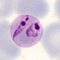
A
-
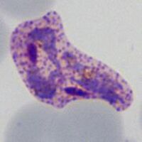
B
-
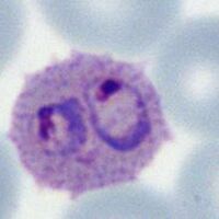
C


Double parasites in: late trophozoite of P.malaria (A) late trophozoite of P.vivax (B) and late trophozoite of P.ovale (C)