Gallery of schizonts: Difference between revisions
From MalariaETC
(Created page with "{{DISPLAYTITLE:<span style="position: absolute; clip: rect(1px 1px 1px 1px); clip: rect(1px, 1px, 1px, 1px);">{{FULLPAGENAME}}</span>}} ---- '''Navigation'''</br> <span style="font-size:90%">>Main Malaria Index''</span></br> <span style="font-size:90%">>>Galleries Index Page''</span></br> <span style="font-size:90%">>>>Current page: '''Gallery of late trophozoites'''</span> ---- <span style="font-size:120%; color:navy">Gallery of Late...") |
No edit summary |
||
| (32 intermediate revisions by the same user not shown) | |||
| Line 4: | Line 4: | ||
<span style="font-size:90%">>[[MalariaETC_Index|Main Malaria Index]]''</span></br> | <span style="font-size:90%">>[[MalariaETC_Index|Main Malaria Index]]''</span></br> | ||
<span style="font-size:90%">>>[[Galleries|Galleries Index Page]]''</span></br> | <span style="font-size:90%">>>[[Galleries|Galleries Index Page]]''</span></br> | ||
<span style="font-size:90%">>>>Current page: '''Gallery of | <span style="font-size:90%">>>>Current page: '''Gallery of schizonts'''</span> | ||
---- | ---- | ||
<span style="font-size: | <span style="font-size:140%; color:navy">'''Gallery of Schizonts'''</br></span> | ||
</br> | </br> | ||
<span style="font-size:90%"> | <span style="font-size:90%">Schizont morphology is variable as they divide their chromatin into separate distinct separate masses (usually a schizont is defined by have more than two masses to distinguish them from rings that have double dots). This morphological variability then continues through successive divisions and maturation. Despite this however, some features such as erythocyte size and shape, added dots, pigment distribution and the number of merozoites present can still be useful (as can the fact that they are rarely seen in ''P.falciparum''.</br></br> | ||
---- | ---- | ||
<span style="font-size:90%">''' ''P.falciparum'' '''</span></br> | <span style="font-size:90%">''' ''P.falciparum'' '''</span></br> | ||
<span style="font-size:90%"> | <span style="font-size:90%">Loose and often "tatty" appearances with 16 or more merozoites and clumped pigment when mature. Rare in blood as they sequester in tissues and circulating form may appear degenerate. Images show mature forms.</br> | ||
<gallery mode="nolines" heights=200px widths=200px> | <gallery mode="nolines" heights=200px widths=200px> | ||
File: | File:PFS1p.jpg|<span style="font-size:80%">Variable merozoite number, clumped pigment</span>|link={{filepath:PFS1p.jpg}} | ||
File: | File:PFS2p.jpg|<span style="font-size:80%">Degenerate small merozoite</span>|link={{filepath:PFS2p.jpg}} | ||
File: | File:PFS3p.jpg|<span style="font-size:80%">Large merozoites</span>|link={{filepath:PFS3p.jpg}} | ||
File: | File:PFS4p.jpg|<span style="font-size:80%">Large merozoites</span>|link={{filepath:PFS4p.jpg}}</gallery>" | ||
---- | ---- | ||
<span style="font-size:95%">''' ''P.vivax'' '''</span></br> | <span style="font-size:95%">''' ''P.vivax'' '''</span></br> | ||
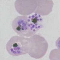
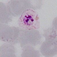
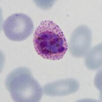
<span style="font-size:90%"> | <span style="font-size:90%">Charactertically large and Schüffner's dots may be seen. The merozoites tend to pack the red cell with numbers up to 16-32 in mature merozoites. Pigment has diffuse distribution. Images show a maturing sequence. | ||
<gallery mode="nolines" heights=200px widths=200px> | <gallery mode="nolines" heights=200px widths=200px> | ||
File: | File:PVS5.jpg|<span style="font-size:80%">Early form with separate chromatin but no individual merozoites</span>|link={{filepath:PVS5.jpg}} | ||
File: | File:PVS1.jpg|<span style="font-size:80%">Similar to previous, note Schüffner's dots</span>|link={{filepath:PVS1.jpg}} | ||
File: | File:PVS2.jpg|<span style="font-size:80%">Maturing form with separate merozoites</span>|link={{filepath:PVS2.jpg}} | ||
File: | File:PVS3.jpg|<span style="font-size:80%">Large mature form, note patchy pigment</span>|link={{filepath:PVS3.jpg}} | ||
</gallery>" | </gallery>" | ||
---- | ---- | ||
<span style="font-size:90%">''' ''P.ovale'' '''</span></br> | <span style="font-size:90%">''' ''P.ovale'' '''</span></br> | ||
<span style="font-size:90%"> | <span style="font-size:90%">Share many features with ''P.vivax'' and may not be easy to distinguish, tend not to be as large with fewer merozoites (up to 16). Ovoid shape and fimbriation of red cells may be present together with James' dots. Pigment has a patchy distribution. | ||
<gallery mode="nolines" heights=200px widths=200px> | <gallery mode="nolines" heights=200px widths=200px> | ||
File: | File:POS1.jpg|<span style="font-size:80%">Early small form note James' dots</span>|link={{filepath:POS1.jpg}} | ||
File: | File:POS3.jpg|<span style="font-size:80%">Early form, merozoites still form a single mass</span>|link={{filepath:POS3.jpg}} | ||
File: | File:POS2.jpg|<span style="font-size:80%">Small mature form with separated merozoites</span>|link={{filepath:POS2.jpg}} | ||
File: | File:POS4.jpg|<span style="font-size:80%">Mature larger form, note ovoid shape</span>|link={{filepath:POS4.jpg}}</gallery>" | ||
---- | ---- | ||
<span style="font-size:90%">''' ''P.malariae'' '''</span></br> | <span style="font-size:90%">''' ''P.malariae'' '''</span></br> | ||
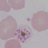
<span style="font-size:90%"> | <span style="font-size:90%">Small and often neat parasites with few merozoites when mature (generally around 8-12). Often there is a central clump of pigment with merozoites arranged around this (daisy head appearance). | ||
<gallery mode="nolines" heights=200px widths=200px> | <gallery mode="nolines" heights=200px widths=200px> | ||
File: | File:PMS1.jpg|<span style="font-size:80%">Early schizont with multiple chromatin dots</span>|link={{filepath:PMS1.jpg}} | ||
File: | File:PMS2.jpg|<span style="font-size:80%">A daisy with merozoites around central pigment</span>|link={{filepath:PMS2.jpg}} | ||
File: | File:PMS3.jpg|<span style="font-size:80%">A less well formed daisy, note small size</span>|link={{filepath:PMS3.jpg}} | ||
File: | File:PMS4.jpg|<span style="font-size:80%">Late form prior to merozoite release</span>|link={{filepath:PMS4.jpg}} | ||
</gallery>" | </gallery>" | ||
---- | ---- | ||
<span style="font-size:90%">''' ''P.knowlesi'' '''</span></br> | <span style="font-size:90%">''' ''P.knowlesi'' '''</span></br> | ||
<span style="font-size:90%"> | <span style="font-size:90%">Similar in appearance to ''P.malariae''. The forms tend to be small with relatively few merozoites present. Pigment tend to form a clump. | ||
<gallery mode="nolines" heights=200px widths=200px> | <gallery mode="nolines" heights=200px widths=200px> | ||
File: | File:PKS3.jpg|<span style="font-size:80%">Early form with few chromatin masses</span>|link={{filepath:PKS3.jpg}} | ||
File: | File:PKS2.jpg|<span style="font-size:80%">Almost a daisy with central clumped pigment</span>|link={{filepath:PKS2.jpg}} | ||
File: | File:PKS1.jpg|<span style="font-size:80%">Prior to release of merozoites</span>|link={{filepath:PKS1.jpg}} | ||
File: | File:PKS4.jpg|<span style="font-size:80%">Immediately before merozoite release</span>|link={{filepath:PKS4.jpg}} | ||
</gallery> | </gallery> | ||
---- | ---- | ||
Latest revision as of 13:29, 17 March 2025
Navigation
>Main Malaria Index
>>Galleries Index Page
>>>Current page: Gallery of schizonts
Gallery of Schizonts
Schizont morphology is variable as they divide their chromatin into separate distinct separate masses (usually a schizont is defined by have more than two masses to distinguish them from rings that have double dots). This morphological variability then continues through successive divisions and maturation. Despite this however, some features such as erythocyte size and shape, added dots, pigment distribution and the number of merozoites present can still be useful (as can the fact that they are rarely seen in P.falciparum.
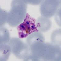
P.falciparum
Loose and often "tatty" appearances with 16 or more merozoites and clumped pigment when mature. Rare in blood as they sequester in tissues and circulating form may appear degenerate. Images show mature forms.
-
Variable merozoite number, clumped pigment
-
Degenerate small merozoite
-

Large merozoites
-
Large merozoites


"
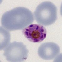
P.vivax
Charactertically large and Schüffner's dots may be seen. The merozoites tend to pack the red cell with numbers up to 16-32 in mature merozoites. Pigment has diffuse distribution. Images show a maturing sequence.
-

Early form with separate chromatin but no individual merozoites
-

Similar to previous, note Schüffner's dots
-

Maturing form with separate merozoites
-

Large mature form, note patchy pigment


"
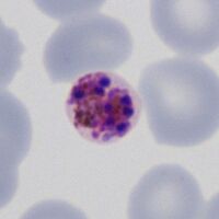
P.ovale
Share many features with P.vivax and may not be easy to distinguish, tend not to be as large with fewer merozoites (up to 16). Ovoid shape and fimbriation of red cells may be present together with James' dots. Pigment has a patchy distribution.
-
Early small form note James' dots
-

Early form, merozoites still form a single mass
-
Small mature form with separated merozoites
-

Mature larger form, note ovoid shape


"
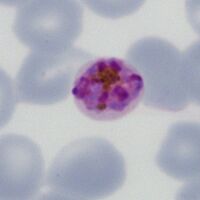
P.malariae
Small and often neat parasites with few merozoites when mature (generally around 8-12). Often there is a central clump of pigment with merozoites arranged around this (daisy head appearance).
-

Early schizont with multiple chromatin dots
-
A daisy with merozoites around central pigment
-
A less well formed daisy, note small size
-
Late form prior to merozoite release


"
P.knowlesi
Similar in appearance to P.malariae. The forms tend to be small with relatively few merozoites present. Pigment tend to form a clump.
-

Early form with few chromatin masses
-

Almost a daisy with central clumped pigment
-

Prior to release of merozoites
-

Immediately before merozoite release


