RDT test: design and function: Difference between revisions
From MalariaETC
No edit summary |
No edit summary |
||
| (37 intermediate revisions by the same user not shown) | |||
| Line 1: | Line 1: | ||
{{DISPLAYTITLE:<span style="position: absolute; clip: rect(1px 1px 1px 1px); clip: rect(1px, 1px, 1px, 1px);">{{FULLPAGENAME}}</span>}} | |||
'''Navigation'''</br> | '''Navigation'''</br> | ||
<span style="font-size:90%">[[Rapid diagnostic tests (RDTs)| | <span style="font-size:90%">[[MalariaETC Index|Main malaria Index]]''</span></br> | ||
<span style="font-size:90%">>>[[Rapid diagnostic tests (RDTs)|RDT main page]]''</span></br> | |||
<span style="font-size:90%">This page: '''How malaria RDTs work'''</span> | |||
---- | ---- | ||
<span style="font-size:160%; color:navy">How Malaria RDT Tests Work</br></span> | |||
---- | |||
{| class="wikitable" style="border-style: solid; border-width: 4px; border-color:light gray" | |||
|colspan="1" style = "font-size:100%; color:black; background: white"|<span style="color:navy></span> | |||
Test formats share common principles, here we illustrate the most simple test format. A "single antigen" test recognising ''P.falciparum''.</br> | <span style="font-size:90%">''Test formats share common principles, here we illustrate the most simple test format. A "single antigen" test recognising ''P.falciparum'' ''.</br></span> | ||
| Line 14: | Line 22: | ||
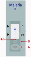
Initially, a buffer is introduced through one window (marked '''A'''); this buffer contains lysing agents which break down the celllar material. The blood sample is then loaded into a second window (marked '''B'''). These two mix to cause lysis of red cells and any malarial parasites they contain. These lysed | <span style="font-size:90%">Initially, a buffer is introduced through one window (marked '''A'''); this buffer contains lysing agents which break down the celllar material. The blood sample is then loaded into a second window (marked '''B'''). These two mix to cause lysis of red cells and any malarial parasites they contain. These lysed constituents then diffuse along the strip to encounter labelled anti-malarial antibody ('''Ab'''). The mixing of antibody and lysed materials allows the antibodies bind to any malaria-specific proteins present in the sample forming immune complexes (antibodies bound to parasite proteins). The mixture of immune complexes, free antibody, and red cells proteins then continues to migrate along the strip driven by excess buffer ('''blue arrow''').</br></span> | ||
<gallery mode="nolines" widths=200px heights= | <gallery mode="nolines" widths=200px heights=200px > | ||
File:1 PF_RDT_pre.jpg|<span style="font-size:90%"></span>|link={{filepath:1 PF_RDT-pre.jpg}} | File:1 PF_RDT_pre.jpg|<span style="font-size:90%"></span>|link={{filepath:1 PF_RDT-pre.jpg}} | ||
</gallery> | </gallery> | ||
| Line 23: | Line 31: | ||
---- | ---- | ||
The process is shown in more detail below: buffer ('''A''') and blood sample ('''B''') enter the strip where red cells and parasites are lysed. The lysed red cells and any parasite antigens then encounter the labelled antibody ('''Ab'''). If malaria antigens are present these labelled antibodies bind to the malarial proteins to form labelled immune-complexes (as shown in the window on the | <span style="font-size:90%">The process is shown in more detail below: buffer ('''A''') and blood sample ('''B''') enter the strip where red cells and parasites are lysed. The lysed red cells and any parasite antigens then encounter the labelled antibody ('''Ab'''). If malaria antigens are present these labelled antibodies bind to the malarial proteins to form labelled immune-complexes (as shown in the window on the image).</span> | ||
<gallery mode="nolines" widths= | <gallery mode="nolines" widths=300px heights=160px> | ||
File:2 RDT_work.jpg|<span style="font-size:90%"></span>|link={{filepath:2 RDT_work.jpg}} | File:2 RDT_work.jpg|<span style="font-size:90%"></span>|link={{filepath:2 RDT_work.jpg}} | ||
</gallery> | </gallery> | ||
Following this initial process, the lysed red cell preparation containing any labelled-immune-complexes then migrates along the strip (red arrow) pushed by the remaining buffer (blue arrow). If the test has been performed correctly the lysed red cells will migrate to the end of the strip. During this process they will first pass over (one or more) test lines ('''T''') then a control control ('''C''') (see below). | <span style="font-size:90%">Following this initial process, the lysed red cell preparation containing any labelled-immune-complexes then migrates along the strip (red arrow) pushed by the remaining buffer (blue arrow). If the test has been performed correctly the lysed red cells will migrate to the end of the strip. During this process they will first pass over (one or more) test lines ('''T''') then a control control ('''C''') (see below).</span> | ||
<gallery mode="nolines" widths= | <gallery mode="nolines" widths=300px heights=160px> | ||
File:2 RDT_work b.jpg|<span style="font-size:90%"></span>|link={{filepath:2 RDT_work b.jpg}} | File:2 RDT_work b.jpg|<span style="font-size:90%"></span>|link={{filepath:2 RDT_work b.jpg}} | ||
</gallery> | </gallery> | ||
| Line 41: | Line 49: | ||
{| class="wikitable" style="border-style: none; border-width: 2px; border-color: gainsboro; color:black" | {| class="wikitable" style="border-style: none; border-width: 2px; border-color: gainsboro; color:black" | ||
|colspan="1" style = "font-size:100%; color:black; background: gainsboro |''' | |colspan="1" style = "font-size:100%; color:black; background: gainsboro |'''"POSITIVE" test result''' | ||
|} | |} | ||
If dye-labelled malaria-antigen/antibody complex is present then it will be "captured" by immobilised anti- | <span style="font-size:90%">If dye-labelled malaria-antigen/antibody complex is present then it will be "captured" by immobilised anti-malarial antibody that also binds the labelled immune complexes. Since there is dye attached to the first antibody this produces a visible line (a positive test line '''T'''). The remaining sample containing labelled antibody only continues to diffuse along the strip and is captured by a second antibody to form a control line ('''C'''). This control line simply indicates that test has been successfully performed.</span> | ||
<gallery mode="nolines" widths= | <gallery mode="nolines" widths=300px heights=260px> | ||
File:3 RDT_work.jpg|<span style="font-size:90%"></span>|link={{filepath:3 RDT_work.jpg}} | File:3 RDT_work.jpg|<span style="font-size:90%"></span>|link={{filepath:3 RDT_work.jpg}} | ||
</gallery> | </gallery> | ||
For this simple test format detecting P.falciparum only, a positive result appears on the test as two visible lines: a '''P.f''' line indicating parasite is present, and a '''C''' showing the test has been correctly performed. Shown below. | <span style="font-size:90%">For this simple test format detecting P.falciparum only, a positive result appears on the test as two visible lines: a '''P.f''' line indicating parasite is present, and a '''C''' showing the test has been correctly performed. Shown below.</span> | ||
<gallery mode="nolines" widths=200px heights= | <gallery mode="nolines" widths=200px heights=200px> | ||
File:3_PF_RDT_pos.jpg|<span style="font-size:90%"></span>|link={{filepath:3_PF_RDT_pos.jpg}} | File:3_PF_RDT_pos.jpg|<span style="font-size:90%"></span>|link={{filepath:3_PF_RDT_pos.jpg}} | ||
</gallery> | </gallery> | ||
| Line 64: | Line 72: | ||
|} | |} | ||
If dye-labelled malaria-antigen/antibody complex is '''not''' present then nothing will be captured by the malaria-specific test line ('''T'''). However, the sample containing labelled antibody that has not bound antigen will still bind to the immobilised antibody that forms control line ('''C''') showing that the test has been successfully performed. | <span style="font-size:90%">If dye-labelled malaria-antigen/antibody complex is '''not''' present then nothing will be captured by the malaria-specific test line ('''T'''). However, the sample containing labelled antibody that has not bound antigen will still bind to the immobilised antibody that forms control line ('''C''') showing that the test has been successfully performed.</span> | ||
<gallery mode="nolines" widths= | <gallery mode="nolines" widths=300px heights=270px> | ||
File:4 RDT_work.jpg|<span style="font-size:90%"></span>|link={{filepath:4 RDT_work.jpg}} | File:4 RDT_work.jpg|<span style="font-size:90%"></span>|link={{filepath:4 RDT_work.jpg}} | ||
</gallery> | </gallery> | ||
Again, for this simple test detecting P.falciparum only, this shows a negative result for ''P.falciparum'' antigens (absent '''P.f''' line), but a positive control line ('''C'''), confirms that the test was correctly performed. Shown below | <span style="font-size:90%">Again, for this simple test detecting P.falciparum only, this shows a negative result for ''P.falciparum'' antigens (absent '''P.f''' line), but a positive control line ('''C'''), confirms that the test was correctly performed.</br>Shown below: </span> | ||
<gallery mode="nolines" widths=200px heights= | <gallery mode="nolines" widths=200px heights=200px> | ||
File:4_PF_RDT_neg.jpg|<span style="font-size:90%"></span>|link={{filepath:4_PF_RDT_neg.jpg}} | File:4_PF_RDT_neg.jpg|<span style="font-size:90%"></span>|link={{filepath:4_PF_RDT_neg.jpg}} | ||
</gallery> | </gallery> | ||
---- | {| class="wikitable" style="border-style: none; border-width: 2px; border-color: gainsboro; color:black" | ||
|colspan="1" style = "font-size:100%; color:black; background: gainsboro |'''Sources''' | |||
|} | |||
* <span style="font-size:90%">[https://www.who.int/teams/global-malaria-programme/case-management/diagnosis/rapid-diagnostic-tests/how-malaria-rdts-work How malaria RDTs work] - World Health Organization (WHO) | |||
* <span style="font-size:90%">[https://www.cdc.gov/dpdx/diagnosticprocedures/blood/antigendetection.html Blood Specimens – Detection of Parasite Antigens] - Centers for Disease Control and Prevention (CDC) | |||
* <span style="font-size:90%">[https://pmc.ncbi.nlm.nih.gov/articles/PMC6278119/ Malaria Rapid Diagnostic Tests: Literary Review] - National Center for Biotechnology Information (NCBI) | |||
Latest revision as of 12:17, 22 March 2025
Navigation
Main malaria Index
>>RDT main page
This page: How malaria RDTs work
How Malaria RDT Tests Work
|
Initially, a buffer is introduced through one window (marked A); this buffer contains lysing agents which break down the celllar material. The blood sample is then loaded into a second window (marked B). These two mix to cause lysis of red cells and any malarial parasites they contain. These lysed constituents then diffuse along the strip to encounter labelled anti-malarial antibody (Ab). The mixing of antibody and lysed materials allows the antibodies bind to any malaria-specific proteins present in the sample forming immune complexes (antibodies bound to parasite proteins). The mixture of immune complexes, free antibody, and red cells proteins then continues to migrate along the strip driven by excess buffer (blue arrow).
The process is shown in more detail below: buffer (A) and blood sample (B) enter the strip where red cells and parasites are lysed. The lysed red cells and any parasite antigens then encounter the labelled antibody (Ab). If malaria antigens are present these labelled antibodies bind to the malarial proteins to form labelled immune-complexes (as shown in the window on the image).
If dye-labelled malaria-antigen/antibody complex is present then it will be "captured" by immobilised anti-malarial antibody that also binds the labelled immune complexes. Since there is dye attached to the first antibody this produces a visible line (a positive test line T). The remaining sample containing labelled antibody only continues to diffuse along the strip and is captured by a second antibody to form a control line (C). This control line simply indicates that test has been successfully performed.
For this simple test format detecting P.falciparum only, a positive result appears on the test as two visible lines: a P.f line indicating parasite is present, and a C showing the test has been correctly performed. Shown below.
If dye-labelled malaria-antigen/antibody complex is not present then nothing will be captured by the malaria-specific test line (T). However, the sample containing labelled antibody that has not bound antigen will still bind to the immobilised antibody that forms control line (C) showing that the test has been successfully performed.
|