Biology of the gametocyte: Difference between revisions
From MalariaETC
No edit summary |
No edit summary |
||
| (2 intermediate revisions by the same user not shown) | |||
| Line 21: | Line 21: | ||
|} | |} | ||
</br> | </br> | ||
<span style="font-size:90%">Gametocytes (like schizonts) are the final mature form of parasites in the blood, consequently they have metabolised most or all of the red cell haemoglobin in order to grow. The red cell therefore simply consistes of membrane/cytoskeleton that surrounds the parasite without any of the red haemoglobin pigment generally with prominent malaria pigment (the metabolised haem molecule containing iron). The appearance of gametocytes however does vary according to species and this can aid morphological species identification</br></br>''' ''P.falciparum'' ''' In this species the gametocytes have a "rod" shape which may be constricted by the remaining red cell membrane to form a curved shape (often described as banana-shaped). | <span style="font-size:90%">Gametocytes (like schizonts) are the final mature form of parasites in the blood, consequently they have metabolised most or all of the red cell haemoglobin in order to grow. The red cell therefore simply consistes of membrane/cytoskeleton that surrounds the parasite without any of the red haemoglobin pigment generally with prominent malaria pigment (the metabolised haem molecule containing iron). The appearance of gametocytes however does vary according to species and this can aid morphological species identification</br></br>''' ''P.falciparum'' '''</br>In this species the gametocytes have a "rod" shape which may be constricted by the remaining red cell membrane to form a curved shape (often described as banana-shaped). | ||
<gallery mode="nolines" widths="150px" heights="150px" > | <gallery mode="nolines" widths="150px" heights="150px" > | ||
File:PFGc.jpg|link={{filepath:PFGc.jpg}} | File:PFGc.jpg|link={{filepath:PFGc.jpg}} | ||
File:PFG4.jpg|link={{filepath:PFG4.jpg}} | File:PFG4.jpg|link={{filepath:PFG4.jpg}} | ||
</gallery></br></br> | </gallery></br></br> | ||
''' ''P.malariae'' ''' | <span style="font-size:90%">''' ''P.malariae'' '''</br>Like other stages in this species the red cell containing the parasite is not enlarged or distorted and may be small. Parasites are typically small and neat with rounded form that do not fill the red cell. | ||
< | |||
<gallery mode="nolines" widths="150px" heights="150px" ></span> | <gallery mode="nolines" widths="150px" heights="150px" ></span> | ||
File:PMGc.jpg|link={{filepath:PMGc.jpg}} | File:PMGc.jpg|link={{filepath:PMGc.jpg}} | ||
File:PMG1.jpg|link={{filepath:PMG1.jpg}} | File:PMG1.jpg|link={{filepath:PMG1.jpg}} | ||
</gallery></br></br> | </gallery></br></br> | ||
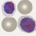
''' ''P.vivax'' ''' | <span style="font-size:90%">''' ''P.vivax'' '''</br>Typically these are very enlarged (significantly larger than normal red cells) often retaining the irregular shape that devlelops in ''P.vivax'' during the trophozoite stage.</span> | ||
< | |||
</br><gallery mode="nolines" widths="150px" heights="150px" > | </br><gallery mode="nolines" widths="150px" heights="150px" > | ||
File:PVGc.jpg|link={{filepath:PVGc.jpg}} | File:PVGc.jpg|link={{filepath:PVGc.jpg}} | ||
File:PVG1.jpg|link={{filepath:PVG1.jpg}} | File:PVG1.jpg|link={{filepath:PVG1.jpg}} | ||
</gallery></br></br> | </gallery></br></br> | ||
''' ''P.ovale'' ''' | <span style="font-size:90%">''' ''P.ovale'' '''</br>Like other species the gametocytes retain the species-specific features acquired during the late trophozoite phase, they may appear sightly enlarged, often ovoid, and have fimbriation.</span> | ||
< | |||
</br><gallery mode="nolines" widths="150px" heights="150px" > | </br><gallery mode="nolines" widths="150px" heights="150px" > | ||
File:POGc.jpg|link={{filepath:POGc.gif}} | File:POGc.jpg|link={{filepath:POGc.gif}} | ||
File:POG4.jpg|link={{filepath:POG4.jpg}} | File:POG4.jpg|link={{filepath:POG4.jpg}} | ||
</gallery></br> | </gallery></br> | ||
''' ''P.knowlesi'' ''' | <span style="font-size:90%">''' ''P.knowlesi'' '''</br>These gametocytes tend to be small like ''P.malariae'' but less neat and well formed, | ||
< | |||
</br><gallery mode="nolines" widths="150px" heights="150px" ></span> | </br><gallery mode="nolines" widths="150px" heights="150px" ></span> | ||
File:PKGc.jpg|link={{filepath:PKGc.jpg}} | File:PKGc.jpg|link={{filepath:PKGc.jpg}} | ||
| Line 54: | Line 50: | ||
|colspan="1" style = "font-size:100%; color:black; background: gainsboro |'''Relevance of gametocytes to clinical biology''' | |colspan="1" style = "font-size:100%; color:black; background: gainsboro |'''Relevance of gametocytes to clinical biology''' | ||
|}</br> | |}</br> | ||
Following treatment of malaria these forms may persist (depending on the nature of the treatment) particularly in the first week but sometimes up to 28 days (unlike the asexual forms that generally disappear more rapidly). | <span style="font-size:90%">Following treatment of malaria these forms may persist (depending on the nature of the treatment) particularly in the first week but sometimes up to 28 days (unlike the asexual forms that generally disappear more rapidly).</br></br> | ||
Latest revision as of 12:02, 20 March 2025
Navigation
>Main Malaria Index
>>Malaria Biology Index
>>>Gametocyte Biology
Biology of the Gametocyte
|
P.malariae P.vivax P.ovale P.knowlesi
Following treatment of malaria these forms may persist (depending on the nature of the treatment) particularly in the first week but sometimes up to 28 days (unlike the asexual forms that generally disappear more rapidly). |