Species identification: summary page: Difference between revisions
From MalariaETC
No edit summary |
No edit summary |
||
| (13 intermediate revisions by the same user not shown) | |||
| Line 7: | Line 7: | ||
|} | |} | ||
</br> | </br> | ||
<span style="font-size:90%">This page presents a broad outline of those species that cause malaria in humans, together with links to more detailed descriptions.</span> | |||
---- | ---- | ||
<div style="width: 95%"> | <div style="width: 95%"> | ||
{| class="wikitable" style="border-style: solid; border-width: 0px; border-color:gray; color:black" | {| class="wikitable" style="border-style: solid; border-width: 0px; border-color:gray; color:black" | ||
|colspan="1" style = "font-size:130%; color:black; background: # | |colspan="1" style = "font-size:130%; color:black; background: #h0ccFF |'''''Plasmodium falciparum''''' | ||
|} | |} | ||
</br> | </br> | ||
<gallery mode="nolines" widths=140px heights=150px> | <gallery mode="nolines" widths=140px heights=150px> | ||
File:PFETc.jpg|<span style="font-size:80%">''Early trophozoite''</span>|link={{filepath: | File:PFETc.jpg|<span style="font-size:80%">''Early trophozoite''</span>|link={{filepath:PFETc.jpg}} | ||
File:PFLTc.jpg|<span style="font-size:80%">Late trophozoite</span>|link={{filepath:PFLTc.jpg}} | File:PFLTc.jpg|<span style="font-size:80%">Late trophozoite</span>|link={{filepath:PFLTc.jpg}} | ||
File:PFSc.jpg|<span style="font-size:80%">Schizont (rare)</span>|link={{filepath:PFSc.jpg}} | File:PFSc.jpg|<span style="font-size:80%">Schizont (rare)</span>|link={{filepath:PFSc.jpg}} | ||
| Line 20: | Line 21: | ||
</gallery> | </gallery> | ||
</br> | </br> | ||
<span style="font-size:90%">'''Summary:''' The ring trophozoites are small and fine with typical accolé forms, multiple parasites per cell, and double dot forms. Red cells aquire characteristic Maurer's dots and clefts in late trophozoites. Schizonts are rarely seen in blood, while gametocytes | <span style="font-size:90%">'''Summary:''' The ring trophozoites are small and fine with typical accolé forms, multiple parasites per cell, and double dot forms. Red cells aquire characteristic Maurer's dots and clefts in late trophozoites. Schizonts are rarely seen in blood, while gametocytes are characteristicaly elongated (and often curved).</span> | ||
</br> | </br> | ||
<span style="font-size:200%">'''→'''</span> [[Plasmodium falciparum: Morphology|<span style="font-size:90%">'''CLICK''' for detailed description</span>]]'' | <span style="font-size:200%">'''→'''</span> [[Plasmodium falciparum: Morphology|<span style="font-size:90%">'''CLICK''' for detailed description</span>]]'' | ||
| Line 27: | Line 28: | ||
<div style="width: 95%"> | <div style="width: 95%"> | ||
{| class="wikitable" style="border-style: solid; border-width: 0px; border-color:gray; color:black" | {| class="wikitable" style="border-style: solid; border-width: 0px; border-color:gray; color:black" | ||
|colspan="1" style = "font-size:130%; color:black; background: # | |colspan="1" style = "font-size:130%; color:black; background: #h0ccFF |'''''Plasmodium vivax''''' | ||
|} | |} | ||
</br> | </br> | ||
| Line 33: | Line 34: | ||
File:PVETc.jpg|<span style="font-size:80%">''Early trophozoite''</span>|link={{filepath:PVETc.jpg}} | File:PVETc.jpg|<span style="font-size:80%">''Early trophozoite''</span>|link={{filepath:PVETc.jpg}} | ||
File:PVLTc.jpg|<span style="font-size:80%">Late trophozoite</span>|link={{filepath:PVLTc.jpg}} | File:PVLTc.jpg|<span style="font-size:80%">Late trophozoite</span>|link={{filepath:PVLTc.jpg}} | ||
File:PVSc.jpg|<span style="font-size:80%">Schizont | File:PVSc.jpg|<span style="font-size:80%">Schizont</span>|link={{filepath:PVSc.jpg}} | ||
File:PVGc.jpg|<span style="font-size:80%">Gametocyte</span>|link={{filepath:PVGc.jpg}} | File:PVGc.jpg|<span style="font-size:80%">Gametocyte</span>|link={{filepath:PVGc.jpg}} | ||
</gallery> | </gallery> | ||
| Line 43: | Line 44: | ||
---- | ---- | ||
<div style="width: 95%"> | <div style="width: 95%"> | ||
{| class="wikitable" style="border-style: solid; border-width: | {| class="wikitable" style="border-style: solid; border-width: 0px; border-color:gray; color:black" | ||
|colspan="1" style = "font-size: | |colspan="1" style = "font-size:130%; color:black; background: #h0ccFF |'''''Plasmodium ovale''''' | ||
|} | |} | ||
</br> | </br> | ||
| Line 50: | Line 51: | ||
File:POETc.jpg|<span style="font-size:80%">''Early trophozoite''</span>|link={{filepath:POETc.jpg}} | File:POETc.jpg|<span style="font-size:80%">''Early trophozoite''</span>|link={{filepath:POETc.jpg}} | ||
File:POLTc.jpg|<span style="font-size:80%">Late trophozoite</span>|link={{filepath:POLTc.jpg}} | File:POLTc.jpg|<span style="font-size:80%">Late trophozoite</span>|link={{filepath:POLTc.jpg}} | ||
File:POSc.jpg|<span style="font-size:80%">Schizont | File:POSc.jpg|<span style="font-size:80%">Schizont</span>|link={{filepath:POSc.jpg}} | ||
File:POGc.jpg|<span style="font-size:80%">Gametocyte</span>|link={{filepath:POGc.jpg}} | File:POGc.jpg|<span style="font-size:80%">Gametocyte</span>|link={{filepath:POGc.jpg}} | ||
</gallery> | </gallery> | ||
| Line 60: | Line 61: | ||
---- | ---- | ||
<div style="width: 95%"> | <div style="width: 95%"> | ||
{| class="wikitable" style="border-style: solid; border-width: | {| class="wikitable" style="border-style: solid; border-width: 0px; border-color:gray; color:black" | ||
|colspan="1" style = "font-size: | |colspan="1" style = "font-size:130%; color:black; background: #h0ccFF |'''''Plasmodium malariae''''' | ||
|} | |} | ||
</br> | </br> | ||
| Line 67: | Line 68: | ||
File:PMETc.jpg|<span style="font-size:80%">''Early trophozoite''</span>|link={{filepath:PMETc.jpg}} | File:PMETc.jpg|<span style="font-size:80%">''Early trophozoite''</span>|link={{filepath:PMETc.jpg}} | ||
File:PMLTc.jpg|<span style="font-size:80%">Late trophozoite</span>|link={{filepath:PMLTc.jpg}} | File:PMLTc.jpg|<span style="font-size:80%">Late trophozoite</span>|link={{filepath:PMLTc.jpg}} | ||
File:PMSc.jpg|<span style="font-size:80%">Schizont | File:PMSc.jpg|<span style="font-size:80%">Schizont</span>|link={{filepath:PMSc.jpg}} | ||
File:PMGc.jpg|<span style="font-size:80%">Gametocyte</span>|link={{filepath:PMGc.jpg}} | File:PMGc.jpg|<span style="font-size:80%">Gametocyte</span>|link={{filepath:PMGc.jpg}} | ||
</gallery> | </gallery> | ||
| Line 77: | Line 78: | ||
---- | ---- | ||
<div style="width: 95%"> | <div style="width: 95%"> | ||
{| class="wikitable" style="border-style: solid; border-width: | {| class="wikitable" style="border-style: solid; border-width: 0px; border-color:gray; color:black" | ||
|colspan="1" style = "font-size: | |colspan="1" style = "font-size:130%; color:black; background: #h0ccFF |'''''Plasmodium knowlesi''''' | ||
|} | |} | ||
</br> | </br> | ||
| Line 84: | Line 85: | ||
File:PKETc.jpg|<span style="font-size:80%">''Early trophozoite''</span>|link={{filepath:PKETc.jpg}} | File:PKETc.jpg|<span style="font-size:80%">''Early trophozoite''</span>|link={{filepath:PKETc.jpg}} | ||
File:PKLTc.jpg|<span style="font-size:80%">Late trophozoite</span>|link={{filepath:PKLTc.jpg}} | File:PKLTc.jpg|<span style="font-size:80%">Late trophozoite</span>|link={{filepath:PKLTc.jpg}} | ||
File:PKSc.jpg|<span style="font-size:80%">Schizont | File:PKSc.jpg|<span style="font-size:80%">Schizont</span>|link={{filepath:PKSc.jpg}} | ||
File:PKGc.jpg|<span style="font-size:80%">Gametocyte</span>|link={{filepath:PKGc.jpg}} | File:PKGc.jpg|<span style="font-size:80%">Gametocyte</span>|link={{filepath:PKGc.jpg}} | ||
</gallery> | </gallery> | ||
Latest revision as of 13:54, 17 March 2025
| Navigation |
| >Main malaria Index |
This page presents a broad outline of those species that cause malaria in humans, together with links to more detailed descriptions.
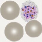
| Plasmodium falciparum |
-

Early trophozoite
-

Late trophozoite
-
Schizont (rare)
-

Gametocyte


Summary: The ring trophozoites are small and fine with typical accolé forms, multiple parasites per cell, and double dot forms. Red cells aquire characteristic Maurer's dots and clefts in late trophozoites. Schizonts are rarely seen in blood, while gametocytes are characteristicaly elongated (and often curved).
→ CLICK for detailed description
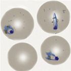
| Plasmodium vivax |
-
Early trophozoite
-

Late trophozoite
-

Schizont
-

Gametocyte


Summary: Large and robust rings may become "amoeboid" in shape during later development. The red cells become increasingly enlarged and distorted and Schüffner's dots are increasingly visible in cytoplams. All forms tend to circulate with large schizont and gametocyte forms present.
→ CLICK for detailed description
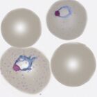
| Plasmodium ovale |
-

Early trophozoite
-

Late trophozoite
-
Schizont
-

Gametocyte


Summary: Trophozites tend to retain a ring appearance but are large and robust. Red cells become moderately enlarged and may have oval shape and/or characteristic fimbriation. Expect James' dots to be easily seen in appropriately stained samples. Cases may be difficult to distinguish morphologically from P.vivax.
→ CLICK for detailed description
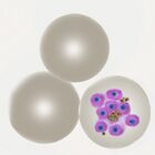
| Plasmodium malariae |
-

Early trophozoite
-

Late trophozoite
-

Schizont
-

Gametocyte


Summary: The species has small ring forms that are less delicate than P.falciparum and which becoming elongated or solid as parasites mature. Red cells are often small retaining their round shape most often with no added dots. Parasite number is often low, but all forms tend to circulate. Look for the characteristic "daisy" schizonts and small round gametocytes
→ CLICK for detailed description
| Plasmodium knowlesi |
-

Early trophozoite
-
Late trophozoite
-

Schizont
-

Gametocyte


Summary: This species has a very limited geographical distribution within S.E Asia. The early trophozoites resemble those of P.falciparum and may have high parasite count, while later rings are more solid or elongated similar to P.malariae, as do schizonts & gametocytes (although less "neat"). Characteristically red cell size is unaffected, although distortion and cytoplasmic dots may be present.
→ CLICK for detailed description